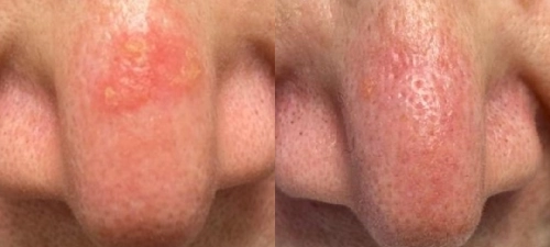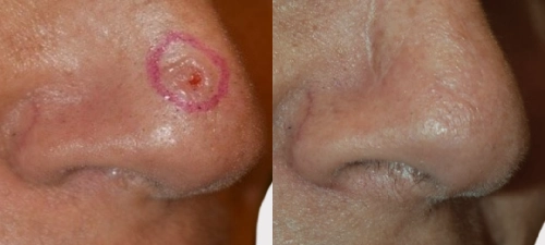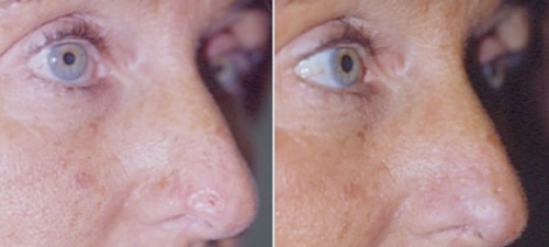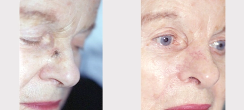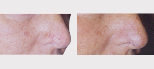
Skin Cancer Diagnosis & Treatment
Dr. Dany Touma has more than 30 years of experience in dermatology and extensive experience in the diagnosis and treatment of skin cancer. His clinical practice includes surgical, laser, and non-surgical approaches selected according to the type, location, extent, and characteristics of each lesion, with consideration for both functional and aesthetic outcomes.
Skin cancer is one of the most common forms of cancer worldwide. It develops when abnormal skin cells grow and multiply in an uncontrolled manner. The three main types are basal cell carcinoma (BCC), squamous cell carcinoma (SCC), and melanoma, each requiring a different approach to diagnosis and treatment.
Understanding the Skin
The skin is the body’s largest organ and provides protection against injury, infection, and dehydration. It consists primarily of three layers:
- Epidermis: the outer protective layer
- Dermis: the deeper connective tissue containing blood vessels, glands, nerves, and hair follicles
- Subcutaneous tissue: the deepest layer, consisting largely of fat and connective tissue
Skin cancer can develop when abnormal cells arise within the skin, often in association with cumulative ultraviolet radiation exposure, genetic factors, or other risk factors.
Main Types of Skin Cancer
Basal Cell Carcinoma (BCC)
Basal cell carcinoma is the most common type of skin cancer. It frequently develops on sun-exposed areas such as the face, scalp, ears, neck, and trunk. BCC usually grows slowly and is unlikely to spread to distant organs, but untreated lesions can invade surrounding tissue and cause significant local damage.
Squamous Cell Carcinoma (SCC)
Squamous cell carcinoma commonly develops on chronically sun-exposed areas, including the face, ears, lips, scalp, and hands. Compared with BCC, some SCCs have a greater potential to invade deeper tissues or spread to lymph nodes and other sites. Early diagnosis and appropriate treatment are important.
Melanoma
Melanoma develops from melanocytes, the pigment-producing cells of the skin. It can occur anywhere on the body and may develop within an existing mole or appear as a new lesion.
Melanoma requires prompt medical assessment because its behaviour can vary according to its type and stage. Early diagnosis is an important factor in treatment planning and prognosis.
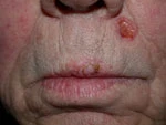
Basal cell Carcinoma of the left upper lip coexisting with Squamous cell carcinoma of the mid lip
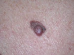
Malignant melanoma
Precancerous Skin Conditions
Actinic Keratoses (Solar Keratoses) are rough, scaly lesions that develop as a result of chronic sun exposure. Some actinic keratoses can progress to squamous cell carcinoma and may therefore require treatment or monitoring.
Depending on the number, location, and characteristics of the lesions, treatment options may include:
- Cryotherapy – freezing the affected tissue
- Laser resurfacing
- Photodynamic therapy (PDT) – using a photosensitizing agent followed by a specific light source
Dr. Dany Touma has contributed to clinical research involving photodynamic therapy for actinic damage and actinic keratoses.
Risk Factors for Skin Cancer
Risk factors may include:
- Fair skin, particularly skin that burns easily
- Light hair or light-coloured eyes
- Significant or cumulative sun exposure
- Use of tanning beds
- A history of severe sunburns
- Multiple or atypical moles (dysplastic nevi)
- A personal or family history of melanoma
- Certain occupational or medical exposures, including some forms of radiation
Skin Cancer Prevention
Sun protection is an important part of reducing cumulative ultraviolet exposure. Measures may include:
- Limiting prolonged direct sun exposure, particularly during periods of intense UV radiation
- Using broad-spectrum sunscreen with SPF 30 or higher
- Wearing protective clothing and a wide-brimmed hat
- Avoiding tanning beds
- Monitoring existing moles and other skin lesions for changes
Warning Signs of Skin Cancer
A dermatological assessment is recommended if you notice:
- A new growth, sore, or patch that does not heal
- A mole that changes in size, shape, or colour
- A lesion that repeatedly bleeds, crusts, or develops persistent itching
- A rapidly changing or unusual pigmented lesion
For pigmented lesions, the ABCDE approach can help identify features that warrant professional assessment:
- A – Asymmetry
- B – Border irregularity
- C – Colour variation
- D – Diameter
- E – Evolution or change over time
The ABCDE criteria are a guide and do not replace a professional skin examination.
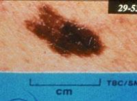
Atypical nevus. Note large size, irregular border and color
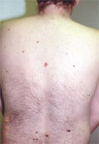
Patient with multiple atypical nevi
Diagnosis
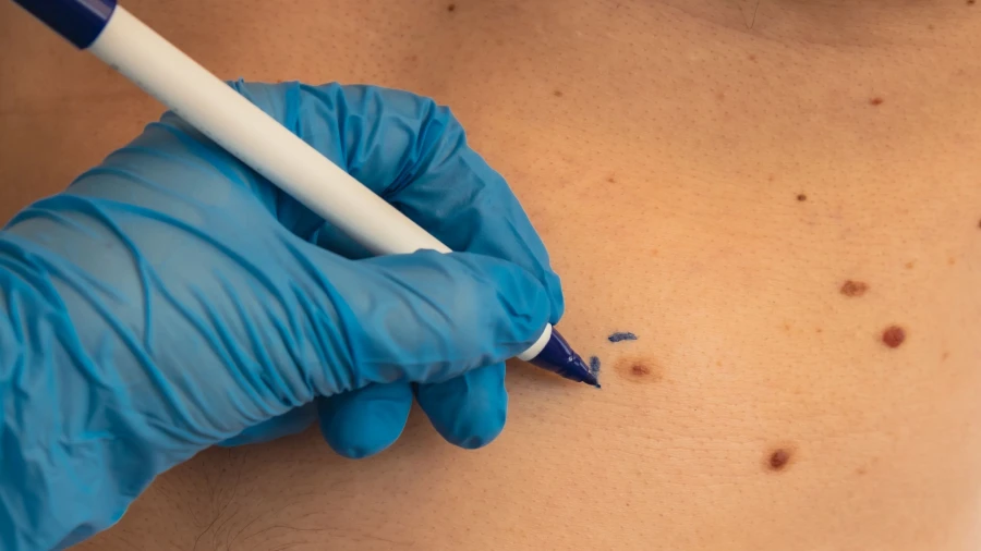
Diagnosis begins with a detailed clinical examination. When a lesion requires further evaluation, a skin biopsy may be performed and examined microscopically to establish the diagnosis and determine relevant pathological characteristics.
Many skin biopsies and minor excisions can be performed under local anaesthesia, depending on the location and extent of the procedure. Post-procedure care and recovery vary according to the type of intervention.
Skin Cancer Treatment
Treatment is selected according to the type and stage of the skin cancer, as well as its size, location, depth, and other clinical characteristics.
Depending on the diagnosis, treatment options may include:
- Excisional surgery – surgical removal of the tumor with an appropriate margin
- Mohs Micrographic Surgery – tissue-sparing surgery with microscopic examination of the surgical margins
- Cryotherapy – controlled freezing of selected lesions.
- Electrodesiccation and curettage – removal and destruction of selected superficial tumors
- Topical therapies – including prescription creams for appropriate superficial lesions
- Radiation therapy – considered for selected cases when clinically indicated
- Other medical or surgical treatments depending on the individual diagnosis
Mohs Micrographic Surgery
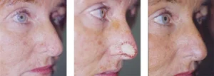
Mohs Micrographic Surgery is a specialized surgical technique particularly considered for selected skin cancers in areas where preservation of surrounding healthy tissue is important, including certain facial tumors and recurrent or complex skin cancers.
The tumor is removed in stages, with the surgical margins examined microscopically during the procedure. Additional tissue is removed only from areas where cancer cells remain, continuing until the examined margins are clear.
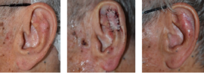
Dr. Dany Touma completed a fellowship in Mohs Micrographic Surgery at Boston University in 1998 and has extensive experience in Mohs surgery and facial skin cancer reconstruction. His practice includes the management of both tumor clearance and reconstruction, with consideration for functional and aesthetic factors.
Melanoma Surgery
Melanoma is primarily treated according to its stage and pathological characteristics. Surgical treatment may involve wider excision of the melanoma and, in selected patients, sentinel lymph node biopsy to evaluate whether cancer cells have spread to regional lymph nodes.
Further treatment and follow-up depend on the individual melanoma diagnosis and stage and may involve a multidisciplinary oncology team.
Follow-Up & Skin Surveillance
Regular dermatological follow-up is important for patients with a history of skin cancer or precancerous lesions. Follow-up recommendations depend on the individual’s diagnosis, treatment history, risk factors, and clinical findings.
Patients may be advised to:
- Perform regular self-examination of their skin
- Attend recommended dermatological follow-up appointments
- Maintain consistent sun-protection habits
- Report new or changing lesions promptly
Early assessment of suspicious skin lesions allows appropriate diagnostic investigation and treatment planning.
Dr. Dany Touma’s Approach to Skin Cancer
With more than 30 years of dermatological experience, Dr. Dany Touma combines clinical assessment, dermatologic surgery, Mohs Micrographic Surgery, laser-based treatments, and other treatment approaches according to the individual skin cancer diagnosis.
Treatment decisions are based on the clinical and pathological characteristics of each case, with attention to cancer management as well as the functional and aesthetic considerations involved in treatment and reconstruction.
If you notice a suspicious mole, spot, growth, or non-healing lesion, a dermatological consultation can help determine whether further examination or biopsy is required.