
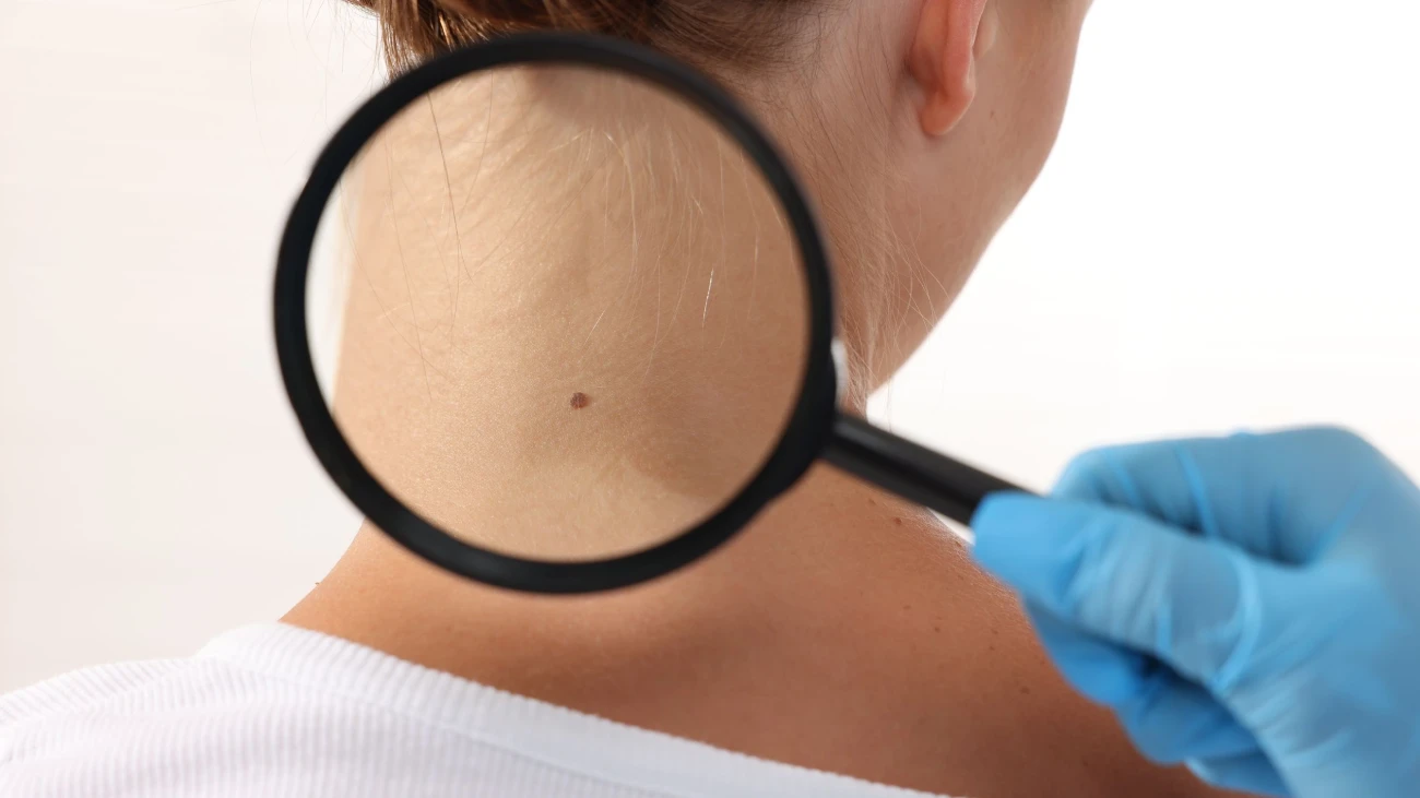
Mole mapping is a structured method of documenting and monitoring moles and other pigmented skin lesions over time. It combines total-body photography and dermoscopic imaging to create a detailed digital record that can be compared during future skin examinations.
Dr. Dany Touma uses the FotoFinder automated dermoscopic system, which combines high-resolution skin imaging with digital documentation and analysis tools to support the clinical assessment and monitoring of pigmented lesions.
Who May Benefit from Mole Mapping?
Mole mapping may be particularly useful for individuals who have:
- Multiple moles or numerous pigmented lesions
- A personal history of skin cancer
- A family history of melanoma or other skin cancers
- Atypical or changing moles
- A need for regular long-term skin surveillance
During a skin examination, suspicious or changing lesions can be assessed using dermoscopy and, when appropriate, further investigated through biopsy or other diagnostic procedures.
Mole mapping is a monitoring tool and does not replace a clinical examination. The frequency of follow-up is determined according to the individual’s skin characteristics, risk factors, and clinical findings.
Regular professional skin checks can help identify changes that may require further assessment and support an individualized approach to long-term skin surveillance.
View this post on Instagram